Sheep brains, like other sheep organs, are much smaller than human brains but have similar features. They can be a valuable addition to your study of anatomy.
See for yourself what the cerebrum, cerebellum, spinal cord, gray and white matter, and other parts of the brain look like with this sheep brain dissection guide! Use this for a high school lab, or just look at the labeled images to get an idea of what the brain looks like.
1. You’ll need a preserved sheep brain for the dissection. Set the brain down so the flatter side, with the white spinal cord at one end, rests on the dissection pan. Notice that the brain has two halves or hemispheres. Can you tell the difference between the cerebrum and the cerebellum? Do the ridges (called gyri) and grooves (sulci) in the tissue look different? How does the surface feel?
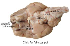
2. Turn the brain over. You’ll probably be able to identify the medulla, pons, midbrain, optic chiasm, and olfactory bulbs. Find the olfactory bulb on each hemisphere. These will be slightly smoother and a different shade than the tissue around them. The olfactory bulbs control the sense of smell. The nerves to the nose are no longer connected, but you can see nubby ends where they were. The nerves to your mouth and lower body are attached to the medulla; the nerves to your eyes are connected to the optic chiasm. Using a magnifying glass, see if you can find some of the nerve stubs.
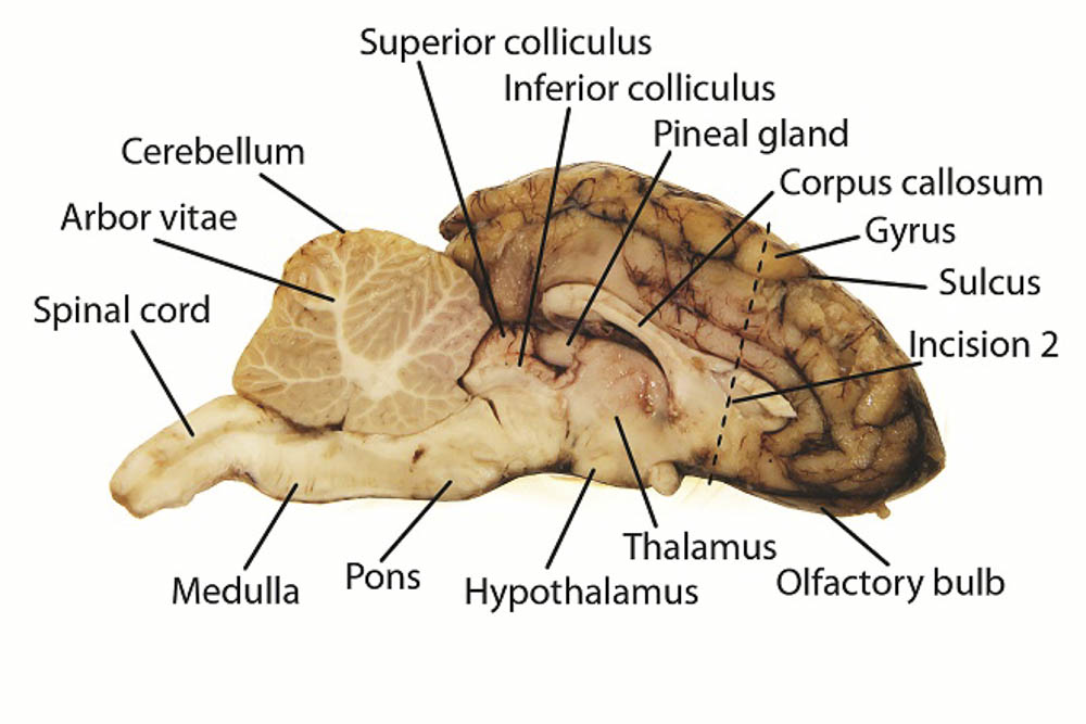
3. Look closely at the inside of the cerebellum. You should see a branching ‘tree’ of lighter tissue surrounded by darker tissue. The branches are white matter, which is made up of nerve axons. The darker tissue is gray matter, which is a collection of nerve cell bodies. You can see gray and white matter in the cerebrum, too, if you cut into a portion of it.
4. You can also use the letter labels on the internal anatomy picture to try to find the following:
Do some more research to find out about these structures in detail.
5. If you have a microscope, slice off a very thin section of the cerebrum and put it on a slide, covering it with a drop of water and a coverslip. Look at it under 100X and 400X magnification. Follow the same procedure with a section of the cerebellum, then compare and contrast the two.
What other users say:
Fun and Educational. We dissected these brains as part of our co-op science class. The kids had a fun time with this hands on experience and it was great to actually be able to locate and see different sections of the brain.
Welcome! Read other Biology / Life Science articles or explore our the rest of the Resource Center which consists of hundreds of free science articles!